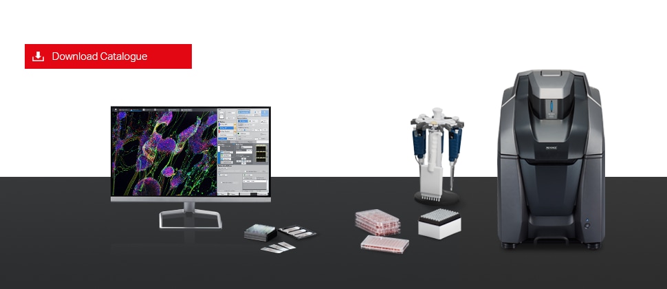
Fluorescence Microscope BZ-X Series
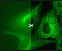
Optical sectioning with structured illumination
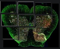
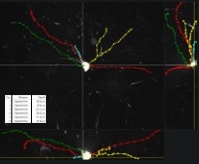
Quickly create high-resolution wide field images
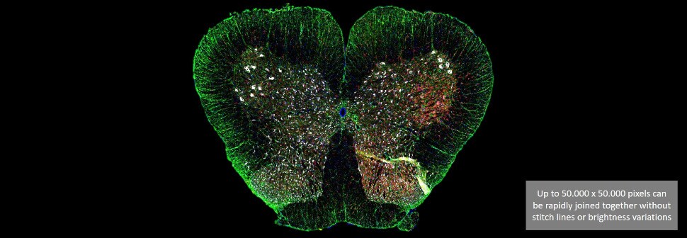 Spinal cord of a rat Courtesy of Professor Tasuku Nishihara, Department of Anesthesia and Perioperative Medicine, Ehime University Graduate School of Medicine
Module selection of the BZ-X model series
-
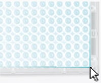
Microplate scanning
-
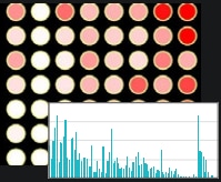
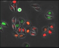
Imaging cytometry
-
Image composition
-
Optical sectioning
-
Live cell imaging
-
Video capture
-
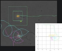
3D measurement and analysis
-
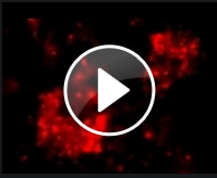
Time lapse analysis
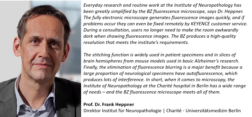
Customer voices
For more information and application examples, download the following catalogue.